In previous literature, several studies have highlighted both harmful and beneficial effects of electromagnetic fields for human beings and, in particular, possible applications for the treatment of some diseases. In this regard, we hypothesize that a possible application of beneficial effects of electromagnetic fields can be represented by irradiating lethal viruses such as SARS-CoV-2 by electromagnetic fields at frequencies close to the natural resonant frequencies of virus capsid and genome, inducing a relevant harmful alteration in its structure. Exposure of weakened virus samples to high-frequency electromagnetic fields at a frequency ranging from 1 to about 40 GHz, for instance, can be carried out by a variable frequency voltage generator and a signal amplifier to detect frequencies at which amplification of Amide and phosphate vibration bands occurs that should induce virus inactivation. FTIR spectroscopy can be used to evaluate the results of exposure. Amide I and II modes and phosphate vibration bands are characteristic of virus capsid and genome, respectively, so that their amplification at a resonant frequency should induce virus damage. Once resonance frequency of SARS-CoV-2 is found for one of its structures, living human spaces can be irradiated by electromagnetic radiation at such frequency to decontaminate not only surfaces but in particular air, contrary to what common disinfectants can do. This methodology could be implemented quite quickly pending the long time required for vaccinations.
INTRODUCTION
A major scientific product concerns the impacts on biological systems as a result of exposure to low and High Frequency (HF) Electromagnetic Fields (EMFs). Such impacts have been highlighted also by FTIR spectroscopy. Indeed, FTIR spectroscopy is capable to supply correct data on the secondary structure of proteins and biological systems. In addition, due to its accurateness, it is possible to quantify the reaction of a biological system to an EMF, such as it has been highlighted by relevant studies published in recent years (Emanuele et al., 2012; Calabro & Magazu, 2014; Calabrò & Magazù, 2015; Calabrò, 2016; Calabrò & Magazù, 2016; Magazù et al., 2016; Calabrò & Magazù, 2017a; Calabrò & Magazù, 2017; Calabrò & Magazù, 2018a; Calabrò & Magazù, 2018b; Calabrò & Magazù, 2019a; Calabrò & Magazu, 2019; El-Bialy, 2021; Panasenko, 2021). This is the main proof why this technique has been selected to hypothesize to carry out this important study for dangerous viruses inactivation like SARS-CoV-2.
This research project aims to quantify by FTIR spectroscopy the effects of EM radiation as a function of frequency on a virus, to detect some resonance frequency at which virus inactivation occurs.
A virus is a simple biological system constituted by a nucleic acid core, the Genome (that can be DNA or RNA) which is surrounded by a protective protein coat, the Capsid, whose main function is to protect the nucleic acid core.
In the case of SARS-CoV-2, in addition to the capsid, it is enveloped by an amphilic lipid bilayer functionalized with viral spike (S) proteins, and its genome is formed by RNA.
Enveloped viruses are most susceptible to environmental stresses, but the human SARS-CoV-2 responsible for Severe Acute Respiratory Syndrome has recently resulted in increasing concern of contact transmission. Once a resonance frequency of SARS-CoV-2 is found for one of its structures, at least, EMF at such frequency can irradiate and decontaminate human spaces (supermarket, hospital, school, cinema, bus, etc.) as usual disinfectants can act on surfaces but cannot inactivate virus dispersed in air due to the continuous influx of people into various environments.
Air Force Research Laboratory (AFRL) is already working on a similar project, even without directing their studies on resonance frequencies.
Instead, the use of EMFs at a specific SARS-CoV-2 natural resonance frequency can preserve humans from harmful effects of EMFs, given that resonant natural frequencies of the human organism should be distinct from that of a virus due to differences between these biological systems.
Furthermore, it can be hypothesized to use a medical device emitting EM radiation at a resonant frequency of SARS-CoV-2, which can be used to irradiate lung area in the human organism in seriously ill patients. In addition, neighboring tissues cannot be damaged by having resonant frequencies different from that of the virus.
MATERIALS AND METHODS
Several biological and chemical techniques have been used up to now to inactivate a virus. Nevertheless, we need a method that can decontaminate human spaces without damaging human health.
Recently it was confirmed how thermal disinfection can reduce coronaviruses (Kampf et al., 2020). Indeed, it was confirmed that heat induces aggregation of the SARS-CoV membrane protein (Lee et al., 2005). Additionally, it was indicated that the nucleo-capsid protein of SARS-CoV is denatured in 10 min at 55 °C (Wang et al., 2004). The authors of this study have already shown by FTIR spectroscopy that EMFs denature proteins of simple organic systems similarly to heat. Indeed, significant comparative enhancement in the β-sheet ingredient considering the α-helix content in proteins secondary structure was observed after exposure to EMFs even at low intensity and this effect was much more evident at high-frequency values (Emanuele et al., 2012; Calabrò & Magazù, 2015; Calabrò & Magazù, 2016; Calabrò & Magazù, 2017; Karpov et al., 2021). Protein denaturation is a necessary condition to inactivate virus capsid so that it can be hypothesized to use HF-EMFs at a resonant frequency to emphasize this effect, that environments can be decontaminated without damaging human organism because of the differences between their natural frequencies.
In literature, many studies have assumed that each biological system has sonant frequencies in the microwave area and that also viruses can be inactivated at resonance frequencies.
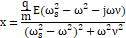
A resonance phenomenon takes place when the frequency of the applied field is close to a fundamental frequency of the system ω = ωs, named natural frequency, giving the maximum value of displacement x. It can be demonstrated that this value is provided by the following equation:
|
|
(1) |
where E is the module of the applied electromagnetic field, ν the modification coefficient, m, and q are the mass and the charge of the particle, ω is the angular frequency of the electromagnetic field and ωs is a characteristic natural frequency of the system.
Following resonance theory, resonance frequencies in SARS-Cov-2 capsid and genome exposed to electromagnetic radiation can be found, searching at what frequency the greatest alterations of vibration bands are observed.
In this regard, several authors have already assumed that viruses may be inactivated using resonance vibrations in the GHz region. In particular, an interesting theoretical study (Yang et al., 2015) schematized the structure of a virus as a nanosphere with a core-shell structure of opposite charge distribution, and the threshold electric field magnitude of the incident microwaves to fracture a virus was at the resonance frequency of 8 GHz.
Experimentally, resonant frequencies in the SARS-CoV-2 envelope exposed to EMF can be found by searching at what frequency the greatest alterations of vibration bands are observed.
In this regard, to test the inhibitory effect of EMFs at resonant frequencies two approaches will be employed: i) the use of HuCoV-229E human coronavirus (Warnes et al., 2015) as a surrogate for the more virulent coronaviruses responsible for COVID-19 (rather than using animal viruses or coronaviruses that initially infect the gastrointestinal tract), to analyze the capsid and RNA genome alteration; ii) the use of Sars-CoV-2 pseudotype to analyze the inhibition of viral binding to the cellular ACE receptor and viral-induced inflammatory response in host cells. The advantage of both HuCoV-229E and Sars-CoV-2 pseudotype virus is that they can be carried out in biosafety level 2 (BSL-2) facilities instead of BSL-3 facilities needed for work with highly pathogenic including new emerging Coronavirus capable to be highly lethal on human.
RESULTS AND DISCUSSION
Viruses structure can be successfully investigated by infrared (IR) spectroscopy, such as it was shown in Santos et al. (2020), in which it was shown that the most specific vibration bands in the mid-IR area of viruses are the Amide I, II, and III vibration bands (due to the viral capsid) and the phosphate bands (due to the genome). SARS-Cov-2 is also enveloped by an amphiphilic lipid bilayer, whose presence can be highlighted in the mid-IR region by symmetric and asymmetric stretching and bending vibration bands of methyl and methylene groups. As a result, the consolidated protocols of the authors of this study concerning the study of biological systems exposed to EMFs by IR spectroscopy can be successfully utilized to investigate the response of SARS-Cov-2 to HF-EMFs, measuring the change in frequency and intensity of its vibration bands as a function of frequency. Hence, the response of SARS-CoV-2 vibration bands to HF-EMF as a function of frequency can be studied. In particular, the maximum level of proteins denaturation (at which SARS-Cov-2 inactivation occurs) should be represented by the largest comparative enhancement in the β-sheet component regarding the α-helix content in the Amide vibration bands, that can be detected by the accurateness of FTIR spectroscopic measurement (Calabro & Magazu, 2014; Calabrò, 2016; Calabrò & Magazù, 2016; Magazù et al., 2016). The corresponding EMF frequency is a resonance frequency.
In addition, viruses’ genome has a high dipole moment, so that a high response of SARS-CoV-2 RNA to an HF-EMF is expected, given that it is free to move as it is not rolled up to the histones like cells. Hence, the genome also would be damaged by using an EMF at a resonance frequency that can be detected by analog spectroscopic measurement of phosphate bands.
The protocol states a 3-hour exposure of weakened samples of SARS-CoV-2 pseudotype to HF-EMF at a frequency ranging from 1 to about 40 GHz, using a variable frequency voltage generator and a signal amplifier placed into an incubator together with virus samples at approximately 10^3 PFU. Unexposed samples will be put into another incubator of the same model and the same physical status. Exposed and unexposed samples will be subjected to FTIR microspectroscopy analysis by Bruker LUMOS and OPUS software for measurement of SARS-Cov-2 vibration bands or any other viruses structure whose study is needed.
Unfortunately, it must be assumed that other contagious and lethal viruses can produce other pandemics in the future and therefore appropriate precautions must be taken. Indeed, we have been able to find from the pandemic for SARS-Cov-2 that it takes a long time for a specific vaccine to be available for the entire population. Instead, the experimental methodology outlined in this paper could be used quite quickly and effectively. In this regard, a project can be carried out aimed at searching the range of natural resonance frequencies of the most important families of viruses that could be cataloged and made available if necessary to direct a specific search for the natural frequencies of a, particularly lethal and contagious virus. Each type of virus would have its particular characteristics and therefore its natural frequencies which should be found specifically using the experimental methodology described here. But in any case, this study should be fast enough as these frequencies should not deviate much from the range of frequencies of his family that would have previously been found.
CONCLUSION
In previous studies, it has been shown that it is possible to quantify the effects of exposure of biological systems to EMFs by FTIR spectroscopy. Such effects should be amplified at particular frequencies that are close to natural resonant frequencies of the biological system. This effect can be applied to dangerous viruses such as SARS-CoV-2. Indeed, resonance frequencies in viruses capsid and genome exposed to electromagnetic radiation can be found by searching at what frequency the greatest alterations of Amide and phosphate vibration bands are observed.
Hence, exposure of weakened samples of a virus to HF-EMF at a frequency ranging from 1 to about 40 GHz, for instance, can be carried out by a variable frequency voltage generator and a signal amplifier to detect frequencies at which increases in the intensity of Amide and phosphate vibration bands occurs.
This method can be used for the inactivation of SARS-CoV-2 or any other virus lethal to humans that may appear in the future. Once the natural resonance frequency of a lethal virus is found for one of its structures, at least, electromagnetic radiation at such frequency can irradiate and decontaminate living human spaces, in particular air, contrary to what common disinfectants can do.
Furthermore, this methodology could be implemented quite quickly pending the long time required for vaccinations.
ACKNOWLEDGMENTS: I am grateful to Prof. Maria Teresa Sciortino (Dep. of Biological and Environmental Sciences, University of Messina, Italy), Prof. Riccardo Ientile, and Dr. Monica Currò (Dep. Bioch. Dent. Sci. Morph. Funct. Image, University of Messina, Italy) for their precious suggestions regarding the use of SARS-CoV-2 pseudotype.
CONFLICT OF INTEREST: None
FINANCIAL SUPPORT: None
ETHICS STATEMENT: None
Air Force Research Laboratory (AFRL): https://www.hill.af.mil/News/Article-Display/Article/2163064/afrl-scientists-investigate-can-microwaves-reduce-viability-of-airborne-coronav/
Calabrò, E. (2016). Competition between hydrogen bonding and protein aggregation in neuronal-like cells under exposure to 50 Hz magnetic field. International Journal of Radiation Biology, 92(7), 395-403.
Calabro, E., & Magazu, S. (2014). Non-thermal effects of microwave oven heating on ground beef meat studied in the mid-infrared region by fourier transform infrared spectroscopy. Spectroscopy Letters, 47(8), 649-656.
Calabrò, E., & Magazù, S. (2015). Fourier self-deconvolution analysis of β-sheet contents in the amide i region of hemoglobin aqueous solutions under exposure to 900 MHz microwaves and bioprotective effectiveness of sugar and salt solutions. Spectroscopy Letters, 48(10), 741-747.
Calabrò, E., & Magazù, S. (2016). Parallel β‐sheet vibration band increases with proteins dipole moment under exposure to 1765 MHz microwaves. Bioelectromagnetics, 37(2), 99-107.
Calabrò, E., & Magazù, S. (2017). Effects of the addition of sodium chloride to a tetrameric protein in water solution during exposure to high frequency electromagnetic field. The Open Biotechnology Journal, 11(1), 72-80.
Calabrò, E., & Magazù, S. (2017a). The α-helix alignment of proteins in water solution toward a high-frequency electromagnetic field: A FTIR spectroscopy study. Electromagnetic Biology and Medicine, 36(3), 279-288.
Calabrò, E., & Magazù, S. (2018a). Direct spectroscopic evidence for competition between thermal molecular agitation and magnetic field in a tetrameric protein in aqueous solution. Physics Letters A, 382(21), 1389-1394.
Calabrò, E., & Magazù, S. (2018b). Resonant interaction between electromagnetic fields and proteins: A possible starting point for the treatment of cancer. Electromagnetic Biology and Medicine, 37(3), 155-168.
Calabrò, E., & Magazù, S. (2019). New perspectives in the treatment of tumor cells by electromagnetic radiation at resonance frequencies in cellular membrane channels. The Open Biotechnology Journal, 13(1), 105-110.
Calabrò, E., & Magazu, S. (2019a). Infrared spectroscopic demonstration of magnetic orientation in SH-SY5Y neuronal-like cells induced by static or 50 Hz magnetic fields. International Journal of Radiation Biology, 95(6), 781-787.
El-Bialy, M., Tawfek, M. A., Hafez, A. M., & Hammad, S. M. (2021). Effect of laser application on pain control during orthodontic tooth movement. Annals of Dental Specialty, 9(1), 62-66.
Emanuele, C., Salvatore, M., & Salvatore, C. (2012). Microwave-induced increase of amide I and amide II vibration bands and modulating functions of sodium-chloride, sucrose and trehalose aqueous solutions: The case study of Haemoglobin. Research Journal of Chemistry and Environment, 16(4), 59-67.
Kampf, G., Voss, A., & Scheithauer, S. (2020). Letter to the Editor- Inactivation of coronaviruses by heat. Journal of Hospital Infection, 105(2), 348-349.
Karpov, V. Y., Zavalishina, S. Y., Bakulina, E. D., Dorontsev, A. V., Gusev, A. V., Fedorova, T. Y., & Okolelova, V. A. (2021). The physiological response of the body to low temperatures. Journal of Biochemical Technology, 12(1), 27-31.
Lee, Y. N., Chen, L. K., Ma, H. C., Yang, H. H., Li, H. P., & Lo, S. Y. (2005). Thermal aggregation of SARS-CoV membrane protein. Journal of Virological Methods, 129(2), 152-161.
Magazù, S., Calabro, E., T Caccamo, M., & Cannuli, A. (2016). The shielding action of disaccharides for typical proteins in aqueous solution against static, 50 Hz and 1800 MHz frequencies electromagnetic fields. Current Chemical Biology, 10(1), 57-64.
Panasenko, S. V., Cheglov, V. P., Ramazanov, I. A., Krasil’nikova, E. A., Stukalova, I. B., & Shelygov, A. V. (2021). Improving the innovative development mechanism of the trade sector. Journal of Advanced Pharmacy Education and Research, 11(1), 141-146.
Santos, M. C., Morais, C. L., Lima, K. M., & Martin, F. L. (2020). Vibrational spectroscopy in protein research toward virus identification: Challenges, new research, and future perspectives. In Vibrational Spectroscopy in Protein Research, Academic Press, 315-335. doi:10.1016/B978-0-12-818610-7.00011-6
Wang, Y., Wu, X., Wang, Y., Li, B., Zhou, H., Yuan, G., Fu, Y., & Luo, Y. (2004). Low stability of nucleocapsid protein in SARS virus. Biochemistry, 43(34), 11103-11108.
Warnes, S. L., Little, Z. R., & Keevil, C. W. (2015). Human coronavirus 229E remains infectious on common touch surface materials. MBio, 6(6), e01697-15. doi:10.1128/mBio.01697-15
Yang, S. C., Lin, H. C., Liu, T. M., Lu, J. T., Hung, W. T., Huang, Y. R., Tsai, Y. C., Kao, C. L., Chen, S. Y., & Sun, C. K. (2015). Efficient structure resonance energy transfer from microwaves to confined acoustic vibrations in viruses. Scientific Reports, 5(1), 18030.
 This work is licensed under a Creative Commons Attribution 4.0 International License.