The production and extraction of enzyme L-aspiraginase produced by some fungus isolates obtained from different soil samples collected from Makkah region, in Kingdom of Saudi Arabia. There were 20 fungal isolates that were recovered from plant rhizosphere in the south and north of Jeddah city, out of which 8 isolates were able to produce L-asparaginase enzyme in high amounts. The higher producers fungal isolates were identified by using DNA sequencing of ITS-1 and ITS-4 primers and deposited in the GenBank nucleotide sequence database under accession numbers as Nothophomagossy piicola LT592943.1, Aspergillus oryzae XR002735719.1, Mucor circinelloides MF356573.1, Aspergillus oryzae HM064501.1, Aspergillus niger DQ915806.1, Rhizopus oryzae JN003654.1, Penicillium sp. KP256500.1, Actinomucor elegansJN887460.1, and Rhizopus oryzae GU126375.1. Nesslerization test showed the difference between the quantities produced by the strains and it was found that the maximum activity of L-asparaginase on the 5th day, was by the isolate AAB1 (1,99 U/min/ml), followed by AGL3(1.94 U/min/ml) and AGL1(1.90 U/min/ml) after six and five days of incubation, respectively.
INTRODUCTION
L-asparaginase was known as L-asparagine amidohydrolase (EC.3.5.1.1). Microbial L-asparaginase has been widely studied in recent years for its potential applications in the pharmaceutical and food industries (Chand et al., 2020; Muneer et al., 2020). L-asparaginase is a useful anti-leukemia enzyme because of its ability in hydrolyzing L-asparagine into aspartic acid and ammonia (Dias & Sato, 2016; Hamid 2021). Asn amino acids are a nutrient critical to the spread and metabolic processes of cancer cells, tumors and Asn depletion leading to the death of leukemia cells (Ali et al., 2016). In the proliferation of leukemic cells, L-asparagine is an important amino acid (Narta et al., 2007). L-asparagine is the single known enzyme used for the degradation of amino acid frequently applied in chemotherapy of cancer (Tork et al., 2019; Sargazi & Taghian, 2020). L-asparaginase is considered as a basis of therapy procedures for severe lymphoblastic leukemia and is applied for retardation stimulation and amplification therapy in the total pediatric treatments and most adult management procedures (Pieters et al., 2011).
There is an extensive variety of microorganisms that can yield this enzyme such as algae, yeast, bacterial, actinomycetes, and fungi, with different properties of enzyme of its clinical therapy applications (Yadav et al., 2014), especially bacteria like Halomonas alkaliantractica (Alzahrani et al., 2019), Bacillus licheniformis (Abdelrazek et al., 2019), Streptomyces fradiae NEAE-82 (Soliman et al., 2020), Sterptomyces sp. (Alzahrani et al., 2020). Certain L-ASNase producing fungal sp., Fusarium equiseti AHMF4 (El-Gendy et al., 2021) Aspergillus oryzae CCT 3940 (Benchamin et al., 2019) Aspergillus terreus (Hassan et al. 2018), Aspergillus flavus (Patro et al., 2014) and Aspergillus terreus (Farag et al., 2015), Penicillium sp. (Cardoso et al., 2020) Penicillium digitatum was screened for producing extracellular L-ASNase (Shrivastava et al., 2016). One of the many producing organisms is the yeast Saccharomyces cerevisiae and also S. marcescens NCIM 2919 (Ghosh et al., 2013).
MATERIALS AND METHODS
Different soil samples were collected from some plants' rhizosphere from Jeddah city from the surface and depth of 10 to 20 cm from different plants rhizosphere, it was serially diluted. Aliquots of 0.1ml of the suspension were evenly spread on Modified Czapek Dox Agar. The plates were incubated at 28C for 72h.
Detection of L-asparagenase activity of fungal isolates (Qualitative assay)
Fungal isolates were appropriately screened for L-asparagenase production capacity by a rapid qualitative screening of the plates using (Czapek yeast extract agar medium) and phenol red (2g in 100ml ethanol) with the final concentration of 0.009% just before pouring the plates. phenol red as an indicator was added to the media, pink color zone around the colonies were reflected L-asparagenase activity (Prakasham et al., 2010).
Quantitative assay of the enzymes by rapid plate method
The potential quantitative estimation of the enzymes in culture filtrate was done by the agar well diffusion method. The fungal Cell-Free Filtrate was harvested by centrifugation at 6000 rpm min-1 for 30 min then filtrate with filter paper. Agar plates were prepared by pouring 20 ml of MCD media into a sterile Petri dish. After plates solidify 7mm wells were punching, using a sterilized cork borer, 100μl of the sample was loaded in the wells, the plates were kept in an upright position at 28±2C˚ for 24 to 48 hrs (Jain et al., 2012).
Following the addition of trichloroacetic acid, by the addition of the enzyme preparation, the blank was started. 0.1ml of the above mixture was taken and added to distilled water of 3.7 ml. After that, 0.2 ml of Nessler’s reagent was also added. Using a visible spectrophotometer, absorbance was measured at 450 nm. Under optimum assay conditions, one international unit of the enzyme (U) was determined the enzyme amount that releases 1 μmol of ammonia from the enzyme. As described by Imada et al. (1973), the enzyme yield was expressed as units/ml.
Formula for the calculation of enzyme activity:
|
|
(1) |
Molecular identification
Genomic DNA extraction
DNA was isolated from fungal isolates using DNeasy® fungi Mini Kit
ITS-u1 and ITS-u4 universal oligonucleotide primers were utilized as described (Cheng et al., 2016). Forward ITS1 (5`TCCGTAGGTGAACCTGCGG3`) and reverse ITS4 (5`TCCTCCGCTTATTGATATGC3`) primers were ordered and synthesized to recover full-length gene.
Database research
The obtained sequencing results were exposed to a BLAST search program (http://www.ddbj.nig.ac.jp/search/blast-j.html) for identification of the similarities with the genes in the gene bank.
RESULTS AND DISCUSSION
Isolation and screening of fungal strains producing L- asparaginase
Based on serial dilution 20 fungal isolates from different soil samples., fungal isolates AAA1, AAA2, and AAA3 were obtained from north Jeddah, while fungal isolates AAB1, AAB2, AAB3, AAB4, and AAB5 were obtained from the samples from east Jeddah, also the fungal isolates AAD1, AAD2, AAD3, AAD4 and AAd5 isolated from south Jeddah. Three fungal isolates AFA1, AFA2and AFA3 were obtained from the samples collected from Khulais, and four fungal isolates AGL1, AGL2, AGL3and AGL4 were isolated from Alkhumra samples.
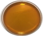
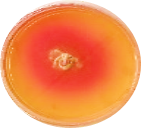
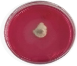
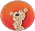
Various fungal strains which were isolated from soil samples were preserved on the modified Czapek dox agar (medium) for further studies. Results in Figure 1 display the collected fungal isolates.
|
L-asparaginase activity |
||
|
|
|
|
|
Control plate |
AAD1 |
AGL3 |
|
|
|
|
|
AAB1 |
AAA1 |
AGL1 |
|
|
|
|
|
AGL4 |
AAD3 |
AAB5 |
|
|
|
|
|
|
AAD4 |
|
|
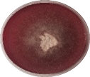
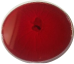
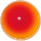
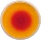
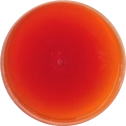
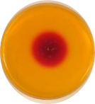
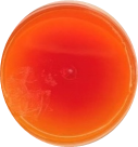
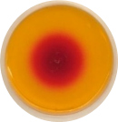
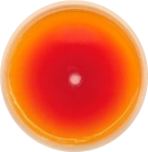
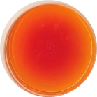
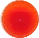
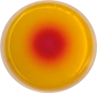
Figure 1. Qualitative assay of L-asparaginase activity of fungal isolates on MCD plates |
||
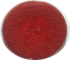
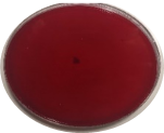
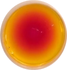
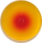
Qualitative assay of L- asparaginase activity of fungal isolates
Fungal isolate's ability for L-asparaginase activity was tested on modified MCD supplemented with L-Asn, as the sole nitrogen source. The initial assay was performed by using phenol red as a pH indicator, Phenol red stain is yellow at acidic pH and changes to pink at alkaline pH, the opposite turn to yellow in the acidic medium. The existence of a pink color region around the colonies on MCD plates with various nitrogen sources was because of the release of the corresponding enzyme.
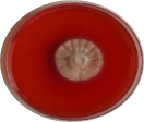
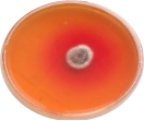
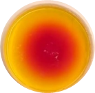
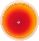
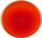
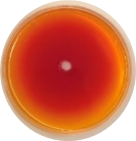
Quantitative assay of L- asparaginase activity in Cell-Free Filtrate of the selected fungal isolates by diffusion method
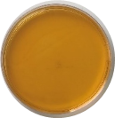
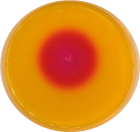
The cell-free filtrate of four selected fungi was evaluated L-asparaginase, production by agar well diffusion assay on solid MCD plates with red phenol, after 24h and 48h of incubation, the measurement of the pink zone in millimeters were recorded based on the diffused of L-asparaginase in the agar.
Results in Figure 2 revealed, the isolates produced different amounts of L-asparaginase after 24h this amounts increased after 48h, the maximum amount of L-asparaginase produced by isolates AAA1, AAB1, AAD3 the diameter of the zone was 58mm, followed by isolate AGL3 the diameter of the zone was 39mm.
|
Fungal Isolates |
12H Control plate |
24H |
Fungal Isolates |
12H |
24H |
|
AGL4 |
|
|
AAA1 |
|
|
|
AGL3 |
|
|
AAB5 |
|
|
|
AAD1 |
|
|
AAB1 |
|
|
|
AGL1 |
|
|
AAD3 |
|
|
|
AAD4 |
|
|
|
|
|
|
Figure 2. Quantitative assay of L-asparaginase activity in Cell-Free Filtrate of the selected fungal isolate by diffusion method. |
|||||
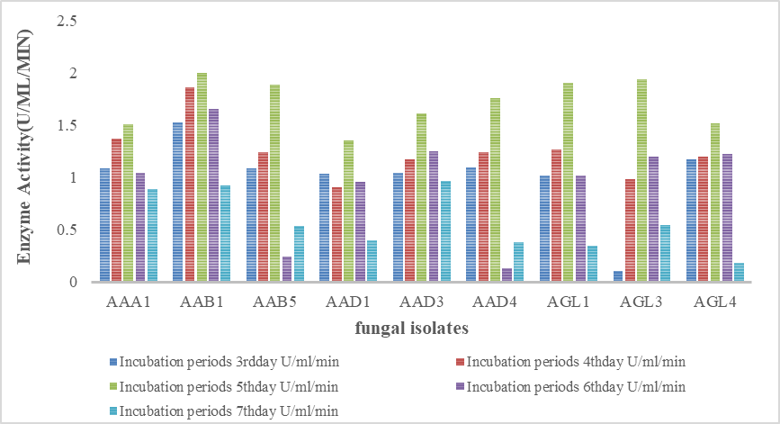
The selected fungal isolates were tested quantitatively for L-asparaginase production and assayed for enzyme activity for 7 days. Results in Figure 3 revealed, the maximum activity of L-asparaginase on the 5th day, was by the isolate AAB1 (1,99 U/min/ml), followed by AGL3(1.94 U/min/ml) and AGL1(1.90 U/min/ml) after six and five days of incubation respectively. The other sex isolates also showed fewer amounts of enzyme in the range of 1.0 to o.6 U/min/ml, the highest amount of each isolate was produced after four days of incubation.
|
|
|
Figure 3. Quantitative assay of L-asparaginase activity produced by the selected fungal isolates by Nessler’s reaction |
Molecular identification of fungal isolates
The fungal isolates were subjected to molecular analysis and identification using DNA sequencing of ITS-u1 and ITS-u4 primers. First, the 750 bp amplicons were separated on 1% agarose gels to prove their accuracy and specificity. Then, the fragments with the molecular sizes representing the ITS-u1 and ITS-u4 primers were purified for sequencing Table 1.
Table 1. DNA sequencing of 18s rRNA using universal primer for fungal isolates
|
Fungal isolate |
Name and Accession of No. of the most related strain in NCBI GenBank |
Identity (%) |
Coverage (%) |
|
|
AAA1 |
Nothophomagossypiicola |
100% |
100% |
|
|
AAB1 |
Aspergillus oryzae |
91% |
100% |
|
|
AAB5 |
Mucor circinelloides |
100% |
100% |
|
|
AAD1 |
HM064501.1 |
Aspergillus oryzae |
100% |
100% |
|
AAD3 |
DQ915806.1 |
Aspergillus niger |
100% |
100% |
|
AAD4 |
JN003654.1 |
Rhizopus oryzae |
100% |
100% |
|
AGL1 |
Penicillium sp. |
100% |
100% |
|
|
AGL3 |
Actinomucorelegans |
100% |
100% |
|
|
AGL4 |
GU126375.1 |
Rhizopus oryzae |
90% |
100% |
Natural products have been the source of the most active ingredients of medicines (Harvey, 2008). Microbial screening programs for the production of enzyme began by isolating the microorganisms and then assayed for enzymatic activity by procedures such as the zone of clearance or zone of color change on agar medium supplemented with appropriate substrate (Balagurunathan et al., 2010; Nagaraju & Ram 2019(. The identification and examination of filamentous fungi which are able to produce extracellular enzymes with biotechnological capacity are considerably important activities (Zambare, 2010; Winter et al., 2021).
The fungal isolates AAA1, AAB1, AAD3 produced different amounts of L-asparaginase after 24h and increased after 48h by isolate AGL3. L-asparaginase's activity in L-asparagine hydrolysis led to aspartic acid and ammonia that transformed red phenol from yellow to pink (El-Naggar & El-Shweihy, 2020). Identical investigations of the production of L-glutaminase by rapid plate assay were found in filamentous fungi by Siddalingeshwara and Lingappa (2010), and with Fusarium oxysporum by Hamed and Al-wasify (2016). Eighteen fungal isolates were obtained from Egyptian marine sponge Aplysinafistularis L-glutaminase had the ability to produced L-glutaminase (Ahmed et al., 2016).
Our results agree with the studies that discovered that variety of fungi produced the anticancer enzyme L-glutaminase. Still, the exclusive enzyme production in the industry has been carried out mainly by Aspergillus species like Aspergillus terreus MTCC 1782 (Varalakshmi & Raju, 2013) may be due to the prevalent nature and non-fastidious nutritional demands of these organisms. Hydrolyses zone surrounding L-ASNase in plate assay was measured to range from 8-13mm for soil bacterial isolates by Devi and Ramanjaneyulu (2016) and (Alzahrani et al., 2019).
About forty-five fungal strains isolated from soil and agricultural remains were examined by using a conventional plate assay process with two indicator stains, phenol red and bromothymol blue (BTB), cleared positive results for L-asparaginase (Doriya & Kumar 2016).
The nine isolates were molecularly analyzed and identified using DNA sequencing of IT1and ITS4 primers. According to Nocker et al. (2004) amplification, ITS1, and ITS4 sequences of fungi can produce 750bp amplicon. The DNA sequences were analyzed using the Blast alignment tools of GenBank. They showed isolates were identified with similarity percentages 99%-100% as: Nothophomagossy piicola LT592943.1, Aspergillus oryzae XR002735719.1, Mucor circinelloides MF356573.1, Aspergillus oryzae HM064501.1, Aspergillus niger DQ915806.1, Rhizopus oryzae JN003654.1, Penicillium sp. KP256500.1, Actinomucor elegansJN887460.1, and Rhizopus oryzae GU126375.1.
About (45%) of fungal isolates showed different activities of L-asparaginase. Aspergillus oryzaeXR-002735719.1(AAB1) showed the maximum L-asparaginase activity (1, 99 U/min/ml) on the 5th day, Assay L-asparaginase activity from an extracellular source by both methods-(Submerged Culture and Nesslerization reaction) (Dhevagi & Poorani, 2006). The extracellular l-asparaginase production from marine actinomycetes was achieved in both submerged and solid-state fermentation conditions (Basha et al., 2009). Also Oliveira et al. (2017) used submerged fermentation where Penicillium chrysogenum had a greater value of L-asparaginase activity (7.00 U/mL). The enzyme activity produced in liquid media reached 8.3 U min.-1 mL-1 (Penicillium sp. T6.2) and 11.4 U min.-1 mL-1 (Fusarium sp.) after 72 hours of cultivation in Bacelar-1 medium (Gonçalves et al., 2016). Mishra (2006) reported that Aspergillus niger showed maximum activity only after 96 hrs of incubation. Many researchers have cleared that 30°C was the optimum temperature for the production of L-asparaginase by Penicillium chrysogenum and Aspergillus terreus (Oliveira et al., 2017; Almisfer et al., 2021). The production of Maximum L-asparaginase (126.67U/min/ml) in medium supplemented with asparagine by Halomonas alkaliantarctica (Al-Zahrani et al., 2019) and was about 33.59 U/Ml in Aspergillus sp. (Doriya & Kumar, 2016). Microorganisms can be easily cultivated and manipulated because they are a good source for enzyme production (Kumar & Sobha, 2012).
CONCLUSION
The current study indicates that the production of L-asparaginase was tested with eight fungal isolates obtained from various soil samples collected from a number of plants in Jeddah, Saudi Arabia. Quantitative and qualitative results showed the high capacity of fungal insulation to produce the enzyme, which is one of the most important enzymes candidates for use in the treatment of cancers. Isolates were molecularly defined by 18S rRNA. It can be concluded from this study that the rooting fungicide activity test has enzyme activity and the extract is expected to be used as therapeutic, industrial and anti-tumor agents.
ACKNOWLEDGMENTS: None
CONFLICT OF INTEREST: None
FINANCIAL SUPPORT: None
ETHICS STATEMENT: None
Abdelrazek, N. A., Elkhatib, W. F., Raafat, M. M., & Aboulwafa, M. M. (2019). Experimental and bioinformatics study for production of L-asparaginase from Bacillus licheniformis: A promising enzyme for medical application. AMB Express, 9(1), 1-16.
Ahmed, A. M. M., Taha, T. M., Abo-Dahab, N. F., & Hassan, F. S. (2016). Process optimization of L-glutaminase production; a tumour inhibitor from marine endophytic isolate Aspergillus sp. ALAA-2000. Journal of Microbial & Biochemical Technology, 8, 256-267.
Ali, U., Naveed, M., Ullah, A., Ali, K., Shah, S. A., Fahad, S., & Mumtaz, A. S. (2016). L-asparaginase as a critical component to combat Acute Lymphoblastic Leukaemia (ALL): A novel approach to target ALL. European Journal of Pharmacology, 771, 199-210. doi:10.1016/j.ejphar.2015,12.023
Almisfer, A. N., Alabbad, H. A., AlHudaithy, H. A. A., Alsultan, N. H., Alobairi, O. K., & Ansari, S. H. (2021). Dental students and dentists’ awareness in handling pediatric patients having systematic diseases in Riyadh. Annals of Dental Specialty, 9(2), 33-38.
Al-Zahrani, N. H., Alzahrani, S. H., & Khayyat, S, K. H. (2020). Isolation and genetic identification of antitumor agent L-Asparaginase Producing Streptomyces sp. Journal of Biochemical Technology, 11(1), 67-73.
Al-Zahrani, S. H. M., Al-Zahrani, N. H., Alamodi, K. H., & Al-Refai, A. (2019). Production of L-Asparaginase as Anticancer Agent by Halomonas alkaliantarctica isolated from marine samples. International Journal of Current Research, 11(04), 3220-3226.
Balagurunathan, R., Radhakrishnan, M., & Somasundaram, S. T. (2010). L-Glutaminase producing actinomycetes from marine sediments–selective isolation, semi quantitative assay and characterization of potential strain. Australian Journal of Basic and Applied Sciences, 4(5), 698-705.
Basha, N. S., Rekha, R., Komala, M., & Ruby, S. (2009). Production of extracellular anti-leukaemic enzyme lasparaginase from marine actinomycetes by solidstate and submerged fermentation: Purification and characterisation. Tropical Journal of Pharmaceutical Research, 8(4), 353-360.
Benchamin, D., Sreejai, R., Sujitha, S., Jensy Roshan, F., Albert, C., & Rishad, K. (2019). Anti-proliferative activity of L-Asparaginase enzyme from fungi on breast cancer. Journal of Pharmacognosy and Phytochemistry, 8(1), 407-410.
Cardoso, S. L., de Freitas, M. M., de Souza, P. M., Homem-de-Mello, M., Silveira, D., Fonseca-Bazzo, Y. M., Filho, E. X., Junior, A. P., & Magalhães, P. O. (2020). Optimization of aqueous two-phase micellar system for partial purification of L-asparaginase from Penicillium sp. grown in wheat bran as agro-industrial residue. Brazilian Journal of Microbiology, 51, 979-988.
Chand, S., Mahajan, R. V., Prasad, J. P., Sahoo, D. K., Mihooliya, K. N., Dhar, M. S., & Sharma, G. (2020). A comprehensive review on microbial l‐asparaginase: Bioprocessing, characterization, and industrial applications. Biotechnology and Applied Biochemistry, 67(4), 619-647.
Devi, A. L., & Ramanjaneyulu, R. (2016). Isolation of L-asparaginase producing microbial strains from soil samples of Telangana and Andhra Pradesh States, India. International Journal of Current Microbiology and Applied Sciences, 5(10), 1105-1113.
Dhevagi, P., & Poorani, E. (2006). Isolation and characterization of L-asparaginase from marine actinomycetes. Indian Journal of Biotechnology, 5, 514-520.
Dias, F. F., & Sato, H. H. (2016). Sequential optimization strategy for maximum l-asparaginase production from Aspergillus oryzae CCT 3940. Biocatalysis and Agricultural Biotechnology, 6, 33-39.
Doriya, K., & Kumar, D. S. (2016). Isolation and screening of L-asparaginase free of glutaminase and urease from fungal sp. 3 Biotech, 6(2), 239.
El-Gendy, M. M. A. A., Awad, M. F., El-Shenawy, F. S., & El-Bondkly, A. M. A. (2021). Production, purification, characterization, antioxidant and antiproliferative activities of extracellular L-asparaginase produced by Fusarium equiseti AHMF4. Saudi Journal of Biological Sciences, 28(4), 2540-2548.
El-Naggar, N. E. A., & El-Shweihy, N. M. (2020). Bioprocess development for L-asparaginase production by Streptomyces rochei, purification and in-vitro efficacy against various human carcinoma cell lines. Scientific Reports, 10(1), 1-21. doi:10.1038/s41598-020-64052-x
Farag, A. M., Hassan, S. W., Beltagy, E. A., & El-Shenawy, M. A. (2015). Optimization of production of anti-tumor l-asparaginase by free and immobilized marine Aspergillus terreus. The Egyptian Journal of Aquatic Research, 41(4), 295-302.
Ghosh, S., Murthy, S., Govindasamy, S., & Chandrasekaran, M. (2013). Optimization of L-asparaginase production by Serratia marcescens (NCIM 2919) under solid state fermentation using coconut oil cake. Sustainable Chemical Processes, 1(1), 1-8.
Gonçalves, A. B., Maia, A. C. F., Rueda, J. A., & Vanzela, A. P. D. F. C. (2016). < b> Fungal production of the anti-leukemic enzyme L-asparaginase: From screening to medium development. Acta Scientiarum. Biological Sciences, 38(4), 387-394.
Hamed, S., & Al-wasify, R. S. (2016). Production and optimization of L-glutaminase from a terrestrial fungal Fusarium oxysporum. International Journal of PharmTech Research, 9(4), 233-241.
Hamid, N. H. M. (2021). Loneliness among the students of faculty of science and arts during the COVID-19. Journal of Organizational Behavior Research, 6(2), 31-45.
Harvey, A. L. (2008). Natural products in drug discovery. Drug Discovery Today, 13(19-20), 894-901.
Hassan, S. W., Farag, A. M., & Beltagy, E. A. (2018). Purification, characterization and anticancer activity of L-asparaginase produced by marine Aspergillus terreus. Journal of Pure and Applied Microbiology, 12(4), 1845-1854.
Jain, R., Zaidi, K. U., Verma, Y., & Saxena, P. (2012). L-asparaginase: A promising enzyme for treatment of acute lymphoblastic leukiemia. People’s Journal of Scientific Research, 5(1), 29-35.
Kumar, D. S., & Sobha, K. (2012). L-asparaginase from microbes: A comprehensive review. Advances in Bioresearch, 3(4), 137-157.
Mishra, A. (2006). Production of L-asparaginase, an anticancer agent, from Aspergillus niger using agricultural waste in solid state fermentation. Applied Biochemistry and Biotechnology, 135(1), 33-42.
Muneer, F., Siddique, M. H., Azeem, F., Rasul, I., Muzammil, S., Zubair, M., Afzal, M., & Nadeem, H. (2020). Microbial L-asparaginase: Purification, characterization and applications. Archives of Microbiology, 202, 967-981.
Nagaraju, K., & Ram, M. R. (2019). Bacillus cereus Rn-6, potential L-Glutaminase producing bacteria from forest soils. Life Science Informatics Publications, 5(4), 134-142.
Narta, U. K., Kanwar, S. S., & Azmi, W. (2007). Pharmacological and clinical evaluation of L-asparaginase in the treatment of leukemia. Critical Reviews in Oncology/Hematology, 61(3), 208-221.
Nocker, A., Lepo, J. E., & Snyder, R. A. (2004). Influence of an oyster reef on development of the microbial heterotrophic community of an estuarine biofilm. Applied and Environmental Microbiology, 70(11), 6834-6845.
Oliveira, M. M., de Araújo, N. K., & Dantas, J. M. D. M. (2017). Evaluation of L-Asparaginase production by microrganisms. In Proceedings of National Bioprocesses Symposium and Enzymatic Hydrolysis of Biomass Symposium (pp. 3-6).
Patro, K. R., Basak, U. C., Mohapatra, A. K., & Gupta, N. (2014). Development of new medium composition for end production of L-asparaginase by Aspergillus f. Journal of Environmental Biology, 35, 295-300.
Pieters, R., Hunger, S. P., Boos, J., Rizzari, C., Silverman, L., Baruchel, A., Goekbuget, N., Schrappe, M., & Pui, C. H. (2011). L‐asparaginase treatment in acute lymphoblastic leukemia: A focus on Erwinia asparaginase. Cancer, 117(2), 238-249.
Prakasham, R. S., Hymavathi, M., Rao, C. S., Arepalli, S. K., Rao, J. V., Kennady, P. K., Nasaruddin, K., Vijayakumar, J. B., & Sarma, P. N. (2010). Evaluation of antineoplastic activity of extracellular asparaginase produced by isolated Bacillus circulans. Applied Biochemistry and Biotechnology, 160(1), 72-80.
Sargazi, M., & Taghian, F. (2020). The effect of royal jelly and exercise on liver enzymes in addicts. Archives of Pharmacy Practice, 11(2), 96-101.
Shrivastava, A., Khan, A. A., Khurshid, M., Kalam, M. A., Jain, S. K., & Singhal, P. K. (2016). Recent developments in l-asparaginase discovery and its potential as anticancer agent. Critical Reviews in Oncology/Hematology, 100, 1-10.
Siddalingeshwara, K. G., & Lingappa, K. (2010). Key fermentation factors for the synthesis of L-asparaginase-an anti tumour agent through ssf methodology. International Journal of Pharmaceutical Sciences, 1(1), 103-112.
Soliman, H. M., El-Naggar, N. E. A., & El-Ewasy, S. M. (2020). Bioprocess Optimization for Enhanced Production of L-asparaginase via Two Model-Based Experimental Designs by Alkaliphilic Streptomyces fradiae NEAE-82. Current Biotechnology, 9(1), 23-37.
Tork, S., Aly, M. M., & Albureikan, M. O. (2019). New tannase-producing lactobacillus Sp. Nrc10: Gene cloning, enzyme purification, and characterization. Pharmacophore, 10(5), 45-56.
Varalakshmi, V., & Raju, K. J. (2013). Optimization of l-asparaginase production by Aspergillus terreus mtcc 1782 using bajra seed flour under solid state fermentation. International Journal of Research in Engineering and Technology, 2(09), 121-129.
Winter, E. A., Wang, L., Tian, M., Litvinova, T. M., Glazkova, I. Y., Winter, K. V., & Zheleznov, A. A. (2021). Employee motivation systems in pharmacies in Japan and China. Journal of Advanced Pharmacy Education and Research, 11(2), 137-145.
Yadav, B., Pemovska, T., Szwajda, A., Kulesskiy, E., Kontro, M., Karjalainen, R., Majumder, M. M., Malani, D., Murumägi, A., Knowles, J., et al. (2014). Quantitative scoring of differential drug sensitivity for individually optimized anticancer therapies. Scientific Reports, 4(1), 1-10.
Zambare, V. (2010). Solid state fermentation of aspergillus oryzae for glucoamylase production on agro residues. International Journal of Life Sciences, 4, 16-25.
 This work is licensed under a Creative Commons Attribution 4.0 International License.