This investigation aimed to study the antioxidant and anti-inflammatory effects of the aqueous extract of Medicgo sativa and copper nanoparticles CuNPs. Biosynthesized CuNPs were characterized by analytical methods. Qualitative phytochemical analysis was carried out by standard protocols. Quantitative Phytochemical analyses were analyzed by using HPLC methods. Moreover, in vivo intraperitoneally acute toxicity testing of nanoparticles was carried up. Results of qualitative phytochemical and quantitative HPLC analysis results revealed that M. sativa contains most of bioactive compounds, especially Naringin, Rutin, and Vanillic Acid compounds with high antioxidant and anti-inflammatory activity. Characterization of CuNPs confirmed the involvement of biological molecules in CuNPs synthesis with size ranged from 19.8 to 92.8 nm. In this study, the intraperitoneal toxicity test showed no mortality and minor behavioral variations up to 20 mg/kg of CuNPs in Wistar rats. We concluded that M. sativa L has potential properties as biocatalyst stabilizers for CuNPs synthesis and MsE-CuNPs revealed good activity as a potent antioxidant, and anti-inflammatory agent. Further in vivo studies are needed to explore them as good therapeutic agents.
INTRODUCTION
The creation of metal and metal oxide nanoparticles (NPs) has significantly improved the biomedical area in recent years in terms of biosensing, imaging, diagnosis, and therapy. The three most often used metals and their oxides are copper, silver, and gold (Cu). (Letchumanan et al., 2021). Due to their unusual physical and chemical characteristics and ease of manufacture, nanoparticles have recently attracted a lot of attention (Derouiche et al., 2022). Today, production of nanoparticles (NPs) using biosynthetictechniques, has been considered as a valuable method with increasing attraction (Keyhani et al., 2018). Cu NPs are effective catalysts that have excellent yields, simple product separation, and may be employed repeatedly. The human body may be harmed by Cu - free ions at the cellular, organ, and systemic levels. Cu ions in living things should therefore be controlled. Inorganic NPs called copper oxides (CuOs) can be produced readily from copper nanoparticles (Cu NPs). Experiments have demonstrated an anti-inflammatory, anti-bacterial, and oxidative stress protective impact for both Cu and CuO NPs, which are both widely utilized as anticancer agents (Ouidad et al., 2020; Derouiche et al., 2022). The ability of NPs to interact with the biological system at the cellular level for numerous reactions and functions is largely responsible for this (Chetehouna et al., 2020). CuO NPs are used in a variety of processes, including catalysis, gas sensing, magnetic phase transitions, antimicrobial activity, and superconductivity (Zahrah, 2022). Current research into the therapeutic effects of plant extracts has revealed several effects of great importance to modern medicine, pharmacy, and industry (Zerrouki & Riaz, 2021). Numerous plant extracts have been used to make copper oxide nanoparticles in large quantities. The copper salts are decreased as a result of the plant extract's production of electrons. In the SEM investigation, the CuNps image was found. Copper oxide nanoparticles are created when phytochemicals combine with copper ions, causing reduction (Siddiqi & Husen, 2020). The nanoparticles, which were bio-synthesised from plant extract, are spherical and less than 90 nm in size (Malik et al., 2022). In this paper, we will choose the Medicago sativa aqueous extract as a base for CuO nanoparticles synthesis (MsE-CuO-NPs); their characterization; anti-oxidant and anti-inflammatory analysis.
MATERIALS AND METHODS
Plant materials and Aqueous extract preparation
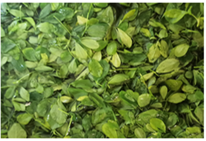
The plant (Medicago sativa) used in this study was purchased from the market. These herbs were mechanically ground into a fine powder before being used. Until the trial started, the Medicago sativa powders were kept at room temperature in airtight containers away from strong light. The aqueous extract was made by mixing 50 g of plant powder dry at 50°C for two hours with 500 ml of distilled water. The mixture was filtered using Whatman paper after 24 hours of maceration at room temperature, and then it was evaporated using a rotary evaporator.
|
|
|
Figure 1. Leave Medicago sativa |
Green synthesis of MsE- Cu Nanoparticles
The appropriate reaction mixture was made for the biogenesis of phyto-copper nanoparticles by adding 2g of copper sulfate into the specified quantity of prepared Medicago sativa aqueous extract (20 ml), and the reaction solutions were combined using a heater-stirred. Both flasks were incubated for 1:30–2 hours in the rotary shaker at 60°C, after which the samples were placed in an electric furnace set to 200°C for 2 hours. Later, using continuous centrifugation (3900 rpm; 10 min; 70°C) with double-distilled water and ethanol, the created phyto-copper nanoparticles (CuNPs) were separated and purified. For additional characterization and bioactivity research, the dried CuNPs were maintained at 60°C (Asemani & Anarjan, 2019).
Characterization of MsE-Cu Nanoparticles
UV-Visible Spectroscopy
UV-visible (UV-Vis) spectroscopy makes it simple to detect the synthesis of copper nanoparticles in Medicago sativa solution. By routinely sampling aliquots (1 mL) of the aqueous component and determining the solution's UV-Vis spectra, the bio-reduction of the cu+ ions in solutions was seen. These aliquots' UV-Vis spectra were observed on a JENWAY 6705 UV-Vis spectrophotometer in the 200-700 nm region operating at a solution of 1 nm as a function of reaction time.
Scanning electron microscope (SEM)
A high-energy electron beam is used to scan a sample using a raster scan pattern in a Scanning electron microscope (SEM). The VERTIV-MODEL 6390 machine was used to perform the SEM and EDX analyses. An amount of the sample was used to create thin films on a carbon-coated copper grid. The extra solution was then blotted off with a piece of paper, and the films on the SEM grid were then dried for five minutes under a mercury lamp.
X-ray diffraction (XRD)
XRD was used to identify the copper nanoparticle's size and makeup. Shimadzu XRD-6000/6100 model with 30 kv, 30 mA, and Cuk a radian at 20 angles were used for this. A quick analytical method that can reveal the dimensions of unit cells is X-ray powder diffraction, which is primarily used to determine the phase of crystalline materials. The average bulk composition of the studied material is found after it has been finely processed. On the copper nanoparticles, the particle or grain size of the particles was measured using Debye Sherrer's equation:
|
D |
(1) |
FTIR spectroscopy
The reduction of Cu ions with a spectrum range of 400–4000 cm-1 was subjected to FTIR analysis to identify the biomolecules that were present in the extract. Here, the sample was dried in a hot air oven and pulverized with KBr to create a pallet before being centrifuged at 3900 rpm for 10 minutes. The pellet was then examined using an FTIR instrument using the Cary 630 model.
Phytochemical analysis, total phenols, and flavonoids compounds
On the aqueous extracts made from the plant using the qualitative characterization method, the phytochemical analysis was done. The Folin-Ciocalteu (FC) method was used to determine the total amount of polyphenols (Boizot & Charpentier, 2006). The procedure given was used to determine the total flavonoids (Dehpour et al., 2009).
Method of chromatographic analysis by HPLC
Before injection, the Medicago sativa extracts were filtered. The following experimental conditions were utilized with an HPLC system, with a detector at = 280 nm for the polyphenols and 360 nm for the flavonoids: other experimental condition: The column used is of length 150 mm and diameter 4.6 mm, the stationary phase C18; Mobile phase: A: acetonitrile; B: 2% glacial acetic acid solution (pH = 2.6); Gradient: 0-5min: 5 % A ; 25-30min: 35% A ; 35-45min: 70%A ; Debit: 0.5 ml / min; Injection volume: 20μL; Temperature: 30 °C.
Antioxidant activity
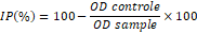
Add 1.25 ml of the buffer solution (0.2 M, PH = 6.6) to 500 l of a sample. 1.25 potassium fericianure should be added. then 20 minutes of incubation at 50 ° C in a water bath. Add 1.25 cc of the 10% aqueous TCA solution after cooling to terminate the reaction. Centrifugation at 3000 rpm for 5 minutes. After that, 1.25 ml of supernatant, 1.25 ml of distilled water, and 250 l of FeCl3 (0.1%) are combined. At 700 nm, the absorbance was calculated in comparison to a blank. Following the computation of the inhibition percentage values in accordance with (Oyaizu., 1986), the following results were obtained by IC50:
|
|
(2) |
Hemolysis assay
A hemolysis assay was performed in accordance with (Vinjamuri et al., 2015). Healthy participants provided 5mL of blood, which was drawn into tubes containing 5.4 mg of EDTA to stop clotting and centrifuged at 1000 rpm for 10 min at 40 °C. The white buffy layer was meticulously aspirated with a pipette after the plasma had been thoroughly removed. The erythrocytes were then washed three more times with 1X PBS, pH 7.4, each time for five minutes. Erythrocytes that had been washed were kept at 4°C and used for the hemolysis assay within 6 hours. The hemolysis assay required 50 L of 10 erythrocytes, which were kept at 4 oC and used within 6 hours. 50 uL of 10 dilutions of erythrocyte suspension (100 uL Erythrocytes suspension: 900 uL 1XPBS) were combined with 100 uL of test samples of Medicago sativa (48 g/mL), 100 uL of 1% SDS, and 100 uL of 1XPBS as positive controls. For 60 minutes, the reaction mixture was incubated in a water bath at 37°C. One mL of the reaction mixture's volume was added by adding 850 L of 1XPB. Finally, the mixture was centrifuged at 300 rpm for 3 minutes, and the amount of hemoglobin in the supernatant was determined at 560 nm using a spectrophotometer. Following are the calculations for percentage of hemolysis inhibition (IH):
|
|
(3) |
Acute toxicity testing
The CuNPs were subjected to an acute toxicity test using Lorke's method. All rats were kept fasting for 12 hours before receiving a single intraperitoneal injection of CuNPs (Control, 10 and 20 mg/kg body weight) into each of the three groups of four (n = 5). There were fifteen (15) rats in total. The rats were watched for 24 hours to track both their behavior and mortality. The department of cellular and molecular biology at El-Oued University's ethics committee examined and gave its blessing to all animal experimentation protocols (approval number: 12 EC/DCMB/FNSL/EU2022).
RESULTS AND DISCUSSION
Phytochemical study of M. sativa aqueous extract
The findings of phytochemical studies indicate that M. sativa aqueous extract is rich in numerous significant chemical components, including flavonoids, phenols, saponins, and terpenoids, but that our plant's extract is made from an alkaloids-reducing ingredient. On the other hand, the findings shown in Table 1 indicated that the aqueous extract of M. sativa was rich in both total phenols and flavonoids.
Table 1. Phytochemical essays for aqueous extract of M. sativa
|
Compounds |
Aqueous extract M. sativa |
|
Flavonoids |
+ |
|
Terpenoids |
+ |
|
Phenols |
+ |
|
Tannins |
+ |
|
Reducing compound |
- |
|
Alkaloids |
- |
|
Saponins |
+ |
|
Polyphenols (mg GAEq/g of extract) |
322.15 ± 22 |
|
Flavonoids (mg QEq/g of extract) |
271.3 ± 17.9 |
(+): Present, (-): Absent
HPLC analysis of M. sativa aqueous extract
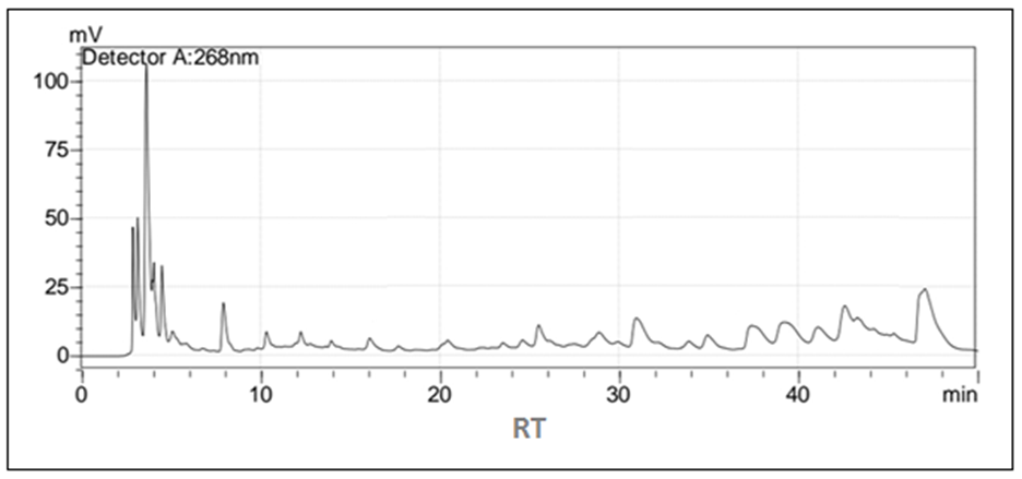
The results of the HPLC chromatographic study demonstrate that M. sativa extract contains a variety of polyphenols in varying amounts, with naringin, rutin, and vanillic acid serving as the primary ingredients (Figure 2 and Table 2).
|
|
|
Figure 2. HPLC chromatogram of phenolic compounds of M. Sativa |
Table 2. Phenolic compounds concentration of M. Sativa aqueous extract
|
Concentration (µg/ml) |
Retention time (min) |
Composition |
|
|
5,29 |
Gallic Acid |
|
1.50 |
13,392 |
Chlorogenic Acid |
|
2.43 |
15,531 |
Vanilic Acid |
|
1.940 |
16,277 |
Caffiec Acid |
|
0.65 |
21,46 |
Vanilin |
|
0.3 |
23,817 |
p-Coumaric Acid |
|
3.87 |
28,37 |
Rutin |
|
12.21 |
34,788 |
Naringin |
|
2.36 |
45,047 |
Quercetin |
Characterization of copper nanoparticles
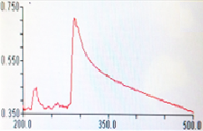
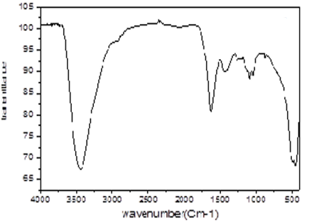
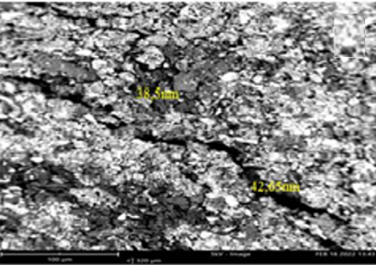
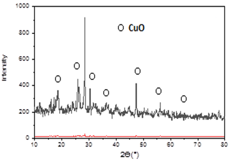
By utilizing a UV-visible spectrophotometer to obtain a spectrum in the visible range of 200 nm-700 nm, the presence of nanoparticles was confirmed (Figure 3a). This investigation revealed an absorbance peak at about 300 that was unique to copper nanoparticles. The chemical make-up of the CuNPs surface was identified and characterized using FTIR spectroscopy. The peak at 3400 cm-1 demonstrated the water and OeH absorption frequency, as can be observed in (Figure 3b). It was done using the SEM (JEOL MODEL 6390) technology to see how big and how shaped the copper nanoparticles were (Figure 3c). The creation of CuNPs and their morphological characteristics in the SEM study showed that the inter-particle spacing was 38.5 nm on average. The copper nanoparticles' shapes showed to be varied. CuO's XRD analysis demonstrated (Figure 3d) the peaks at 18.13°, 19.87°, 22.28°, 25.37°, 25.89°, 26.34°, 36.44°, 78.09° that symbolized by (O).
|
|
|
a) |
|
|
|
b) |
|
|
|
c) |
|
|
|
d) |
|
Figure 3. Characterization of copper nanoparticles UV-VIS spectrum (a), FTIR spectrum (b), SEM image (c), and X-ray diffraction diffractograms (d). |
Antioxidant activity
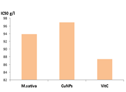
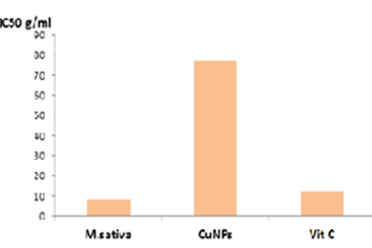
Figure 4a reports on the FRAP assay's outcomes. Vitamin C showed the strongest inhibitory efficacy, up to a maximum of 90.68% at a concentration of 0.7 mg/mL, while M. sativa Reported a concentration of 93.85% at 0.8 mg/ml and 98.63% at 0.8 mg/ml for Cu NPs. The amount of M. sativa extract needed to inhibit free radicals by 50% (IC50) was 17.32 g/ml. IC50 values for M. sativa aqueous extract with regression coefficients (R2=0.99). High regression coefficients and IC50 values for the common vitamin C were 4.12 g/mL.
|
|
|
a) |
|
|
|
b) |
|
Figure 4. IC50 values for M. sativa aqueous extract, CuNPs, and Vitamin C, a for FRAP test and b for hemolysis test |
Hemolysis protection
Human blood erythrocyte membrane dehydration and delayed proton equilibria caused by phosphate buffer (XPBS, pH= 7.4) result in membrane degradation and eventual hemolysis. The antihemolytic properties of the different concentrations (0.2-1 mg/mL) of M. sativa. The effects of M. sativa and CuNPs on human blood erythrocytes were shown in Figure 4b. The maximum antihemolysis activity of M. sativa's aqueous extract was 8.60% at a concentration of 1 mg/mL, while CuNPs' antihemolysis activity peaked at 91.11% at the same dose. Contrary to popular belief, antihemolysis action increased with decreasing CuNP content at different concentrations. On the other hand, we observed the exact opposite: at different concentrations of M. sativa aqueous extract, the antihemolysis activity increased with increasing concentration. Aqueous extract of M. sativa has anti-hemolysis activity with a high regression coefficient (R2 = 0.9184), and regression was also present in CuNPs (R2 = 0.5692).
Acute toxicity study
The test's findings (Table 3) demonstrated that physiological parameters were normal throughout the experiment, which involved treating albino Wister rats with different doses of CuNPs (0, 10, and 20 mg/kg) to affect their eyes, sleep, movement, and diarrhea. Additionally, no unusual symptoms or side effects appeared, and no fatality cases were reported before 14 days. These outcomes supported research demonstrating the medicinal and safe uses of these environmentally friendly Cu nanoparticles.
Table 3. Acute toxicity test of CuNPs on physiological parameters of Wistar albino rats
|
Parameters |
0 h |
3 h |
24 h |
Day- 7 |
Day-14 |
|||||
|
Control |
Test |
Control |
Test |
Control |
Test |
Control |
Test |
Control |
Test |
|
|
Dead rats |
0 |
0 |
0 |
0 |
0 |
0 |
0 |
0 |
0 |
0 |
|
Eyes |
N |
N |
N |
N |
N |
N |
N |
N |
N |
N |
|
Sleep |
N |
N |
N |
N |
N |
N |
N |
N |
N |
N |
|
Movement |
N |
N |
N |
N |
N |
N |
N |
N |
N |
N |
|
Diarrhea |
N |
N |
N |
N |
N |
N |
N |
N |
N |
N |
N: Normal
The findings of phytochemical investigations indicate that M. sativa's aqueous extract contains a variety of significant secondary metabolic compounds, such as flavonoids, phenols, carbohydrates, saponosides, and terpenoids, but does not include tannins or alkaloids. Bora and Sharma (2012) found that the phytochemical finding was identical to our result. The biological features of secondary plant metabolites include antioxidant activity, antibacterial activity, antidiabetic activity, anti-inflammatory activity (Chetehouna et al., 2021), hypolipidemic activity, anxiolytic activity, dermatological activity, immunological activity, and cytotoxic activity (Ali Esmail et al., 2021). According to a prior study, a lot of focus is placed on the effects of saponins found in alfalfa. Studies using macaques failed to find any evidence of the harmful effect of alfalfa saponins (Raeeszadeh et al., 2021; Ahmed et al., 2022). Saponins have been linked to increased production of bile, gastric, pancreatic, and intestinal fluids as well as a reduction in total cholesterol levels (Samir et al., 2020; Romero-Martínez et al., 2021). The research showed that they may exhibit antimycotic and antibacterial activities depending on their chemical structure, mostly against certain yeast harmful to humans. As a result, they might potentially stimulate the immune system. Alfalfa spawning was found to be most active against Gram-positive bacteria like Staphylococcus aureus and Enterococcus faecalis. The in-vitro tests proved their effectiveness as anticancer drugs and established their capacity to stop the growth of human leukemia cell lines. The amount of total phenolic and flavonoid components in the aqueous M. sativa extract is larger than the amounts noted by Karimi et al. (2013), indicating that the concentration is based on the sample's features and the extraction process used.
According to our research, the combination turned black when CuSO4 was added to green-synthesized CuNps made from M. sativa extract. This, in our opinion, points to the production of CuO. Furthermore, that is comparable to a study (Ouidad et al., 2020), The phenolic components, flavonoids, saponins, carbohydrates, and nargenin that are abundant in our plant extract have antioxidant characteristics and biological reductivity in the creation of nanoparticles. We propose that hydrogen ions are divorced during net diversion because it changes the copper ion status to copper condition while also oxidizing it to generate nanoparticles (Bezza et al., 2020) Compared to the following research, for the enol form of flavonoids to the quito shape, reducing copper ions and the synthesis of CuNPs nanoparticles (Mroczek-Sosnowska et al., 2013; Istyagina-Eliseeva et al., 2022).
One of the crucial methods for confirming the initial presence of metal nanoparticles in a liquid media is UV-Vis spectroscopy (Djouadi & Derouiche, 2021). The UV-Vis spectrophotometer was used to further describe the color shift that indicated the presence of copper nanoparticles, and readings were taken at a specific temperature to detect it. The UV-Vis spectra of copper nanoparticle production utilizing Medicago sativa nanoparticle extract are visible in the absorption peaks. Peaks in the 200–700 nm spectral lines in the UV–visible spectra were seen as a result of copper colloid's surface plasma resonance at various temperatures. Around 435 nm, there were 300 strong SPR bands. Our findings support the observations that copper nanoparticle formation occurs. Results regarding the FTIR spectra of CuNPs showed peaks at 3500, 1600, and. The band at 3452 cm 1 is said to correspond to the polymeric OH stretching mode. Amido and open chain amino (C N) are represented by the absorption at 1629 cm 1. According to methyl C-H, 1388 cm1 is the Siam bend. This evidence suggests that the ability to arrange and regulate CuNPs in an aqueous media could be performed by protein particles (Popa et al., 2022). The crystallinity of the synthesized particle structures were revealed by X-ray diffraction (XRD) analysis, which revealed the existence of several sharply pronounced reflections (Shutov et al., 2017). The XRD methodology demonstrated in the work by Emmalai and Velmurugan (2015), which utilized Azadirachta indica (L.) for the synthesis of CuO nanoparticles, confirms the environmentally acceptable approach of nanoparticle synthesis. The SEM scan revealed nanoparticles with diameters between 38.5 and 65.5 nm that were substantially spherical. Memo et al., 2020, who create copper nanoparticles (CuNPs) using an aqueous leaf extract of Ziziphus mauritiana L., reported a similar result. Copper nanoparticles demonstrate extraordinary performance as antibacterial and antimicrobial agents (Xinzhen et al., 2021). Because M. sativa plants contain a lot of phenolic compounds, flavonoids, and saponins, all of which have high antioxidant potential, their extract has a strong antioxidant impact, according to studies on anti-oxidant activity (Michalak, 2022). According to FRAP experiments, they are powerful free radical scavengers because they reduce iron by proportionally donating hydrogen to the extract concentration (Rafiska et al., 2017). Free radicals are known to quietly contribute to the oxidation of unsaturated fat in foods, but they are also combated by phenolic compounds, flavonoids, and saponins. Our extract has contributed to antioxidant activity because it contains a lot of these components. As CuNPs were used to treat the albino Wister rats' eyes, sleep, movement, and diarrhea during the study period, the results of the acute toxicity test revealed that there were normal physiological parameters during that time. No unusual symptoms, negative side effects, or cases of mortality were recorded before 14 days, either. The uses of these green-synthesized Cu nanoparticles for both medicinal and safe purposes were demonstrated by this study's results, which were confirmed (Atoussi et al., 2020).
CONCLUSION
In conclusion, we were successful in synthesizing high-quality, pure monodispersed CuO nanoparticles using alfalfa extract. Additionally, both the created CuNPs and the alfalfa extract were able to provide powerful antioxidant and anti-inflammatory capabilities.
ACKNOWLEDGMENTS: This work was supported by the research project D01N01UN390120210002 funded by the ministry of higher education, Algeria.
CONFLICT OF INTEREST: None
FINANCIAL SUPPORT: None
ETHICS STATEMENT: The study had the approval of the ethics Committee of the El-Oued University (12 EC/CMB/FNSL/EU2022).
Ahmed, H. K., Eisa, I. M., Abdallah, E. I., Hamouda, D. G., Omer, A. E., & Eltayeb, L. B. (2022). Appraisal of biosafety measures in governmental medical laboratory personnel: Knowledge, attitude, practice (KAP) study. Journal of Biochemical Technology, 13(3), 13-18.
Ali-Esmail, A., Hanaa, S. K., Hussein-Ali, A., Alqahtani, A. M., Batiha, G. E., & Jafari-Sales, A. (2021). A review on Medicago sativa: A potential medicinal plant. International Journal of Pharmaceutical and Biological Science Archive, 1(2), 22-33.
Asemani, M., & Anarjan, N. (2019). Green synthesis of copper oxide nanoparticles using Juglans regia leaf extract and assessment of their physico-chemical and biological properties. Green Processing and Synthesis, 8(1), 557-567.
Atoussi, O., Chetehouna, S., & Derouiche, S. (2020). Biological properties and acute toxicity study of copper oxide nanoparticles prepared by aqueous leaves extract of portulaca oleracea (L). Asian Journal of Pharmaceutical Research, 10(2), 89-94.
Bezza, F. A., Tichapondwa, S. M., & Chirwa, E. (2020). Fabrication of monodispersed copper oxide nanoparticles with potential application as antimicrobial agents. Scientific Reports, 10(1), 1-18. doi:10.1038/s41598-020-73497-z
Boizot, N., & Charpentier, J. P. (2006). Méthode rapide d’évaluation du contenu en composés phénoliques des organes d’un arbre forestier. Cahier des Techniques de l'INRA, 79-82.
Bora, K. S., & Sharma, A. (2011). Evaluation of antioxidant and cerebroprotective effect of medicago sativa Linn. against ischemia and reperfusion insult. Evidence-Based Complementary and Alternative Medicine, 2011. doi:10.1093/ecam/neq019
Chetehouna, S., Atoussi, O., & Derouiche, S. (2020). Biological activity and toxicological profile of zinc oxide nanoparticles synthesized by Portulaca oleracea (L) leaves extract. Advances in Nanomedicine and Nanotechnology Research, 2(2), 125-133.
Chetehouna, S., Atoussi, O., & Derouiche, S. (2021). An overview of Portulaca oleracea: Phytochemistry and pharmacological activities. International Journal of Pharmaceutical and Clinical Research, 3(1), 15-18.
Dehpour, A. A., Ebrahimzadeh, M. A., Fazel, N. S., & Mohammad, N. S. (2009). Antioxidant activity of the methanol extract of Ferula assafoetida and its essential oil composition. Grasas y Aceites, 60(4), 405-412.
Derouiche, S., Laib, I., & Zeribit, W. (2022). Protective effect of zinc acetate and Zinc - Aristolochia longa extract nanoparticles against nickel-induced acute liver and kidney injury in rats. Annals of the Romanian Society for Cell Biology, 26(1), 307-316.
Djouadi, A., & Derouiche, S. (2021). Spinach mediated synthesis of zinc oxide nanoparticles: Characterization, In vitro biological activities study and in vivo acute toxicity evaluation. Current Research in Green and Sustainable Chemistry, 4, 100214.
Istyagina-Eliseeva, E., Myagkova, S., & Litvinov, S. (2022). Expansion of grant-based (Scholarship Programs) globalization in education. Journal of Organizational Behavior Research, 7(2), 48-59.
Karimi, E., Oskoueian, E., Oskoueian, A., Omidvar, V., Hendra, R., & Nazeran, H. (2013). Insight into the functional and medicinal properties of Medicago sativa (Alfalfa) leaves extract. Journal of Medicinal Plants Research 7(7), 290-297.
Keyhani, A., Mahmoudvand, H., Shakibaie, M., Tavakoli Kareshk, A., & Nejati, J. (2018). Histopathological and toxicological study of selenium nanoparticles in BALB/C Mice. Entomolgy Applied Science Letter, 5(1), 31-35.
Letchumanan, D., Sok, S. P., Ibrahim, S., Nagoor, N. H., & Arshad, N. M. (2021). Plant-based biosynthesis of copper/copper oxide nanoparticles: An update on their applications in biomedicine, mechanisms, and toxicity. Biomolecules, 11(4), 564. doi:10.3390/biom11040564
Malik, M., Iqbal, M. A., Malik, M., Raza, M. A., Shahid, W., Choi, J. R., & Pham, P. V. (2022). Biosynthesis and characterizations of silver nanoparticles from annona squamosa leaf and fruit extracts for size-dependent biomedical applications. Nanomaterials, 12(4), 616.
Michalak, M. (2022). Plant-derived antioxidants: Significance in skin health and the ageing process. International Journal of Molecular Sciences, 23(2), 585.
Mroczek-sosnowska, N., Batorska, M., Ukasiewicz, M., Wnuk, A., Sawosz, E. W. A., Jaworski, S. A., & Niemiec, J. A. N. (2013). Effect of nanoparticles of copper and copper sulfate administered in ovo on hematological and biochemical blood markers of broiler chickens. Annals of Warsaw University of Life Sciences-SGGW. Animal Science, 149(52), 141-149.
Ouidad, A., Sara, C., & Samir, D. (2020). Biological properties and acute toxicity study of copper oxide nanoparticles prepared by aqueous leaves extract of portulaca oleracea (L). Asian Journal of Pharmaceutical Research, 10(2), 89-94. doi:10.5958/2231-5691.2020.00017.9
Oyaizu, M. (1986). Studies on products of browning reaction antioxidative activities of products of browning reaction prepared from glucosamine. The Japanese Journal of Nutrition and Dietetics, 44(6), 307-315.
Popa, C., Petrus, M., & Bratu, A. M. (2022). Alfalfa (Medicago sativa) sprouts respiratory responses to cadmium stress using IR LPAS. Molecules, 27(6), 1891.
Raeeszadeh, M., Moradi, M., Ayar, P., & Akbari, A. (2021). The antioxidant effect of Medicago sativa L.(alfalfa) ethanolic extract against mercury chloride (HgCl2) toxicity in rat liver and kidney: An in vitro and in vivo study. Evidence-Based Complementary and Alternative Medicine, 2021. doi:10.1155/2021/8388002
Rafińska, K., Pomastowski, P., Wrona, O., Górecki, R., & Buszewski, B. (2017). Medicago sativa as a source of secondary metabolites for agriculture and pharmaceutical industry. Phytochemistry Letters, 20, 520-539.
Romero-Martínez, N., Ramos-Zambrano, E., Osorio-Ruiz, A., & Martínez-Ayala, A. L. (2021). Main mechanisms of action of policosanol in animal and plant cells. International Journal of Pharmaceutical Research and Allied Sciences, 10(2), 10-20.
Samir, D., Yousraa-Imane, G., & Islam, B. (2020). Characterization and acute toxicity evaluation of the MgO nanoparticles synthesized from aqueous leaf extract of ocimum basilicum L. Algerian Journal of Biosciences, 1(1), 1-6.
Shutov, D. A., Rybkin, V. V., Ivanov, A. N., & Smirnova, K. V. (2017). Synthesis of zinc oxide powders in plasma–solution systems. High Energy Chemistry, 51(1), 65-69.
Siddiqi, K. S., & Husen, A. (2020). Current status of plant metabolite-based fabrication of copper/copper oxide nanoparticles and their applications: A review. Biomaterials Research, 24(1), 1-15.
Vinjamuri, S., Afshan, S., Shekar, S., & Saraswathi, V. J. (2015). Evaluation of hemolytic activity, ATPase inhibitory activity and antitumor activity of TLC extract of lemon grass (Cymbpogon citratus). International Journal of Pharmacognosy and Phytochemical Research, 7(4), 785-788.
Xinzhen, F., L’Hocine, Y., & Edward, S. (2021). Antimicrobial properties of the Ag, Cu nanoparticle system. Biology, 10(2), 137. doi:10.3390/biology10020137
Zahrah, A. (2022). Green synthesis of copper oxide nanoparticles CuO NPs from eucalyptus globoulus leaf extract: adsorption and design of experiments. Arabian Journal of Chemistry, 15(5), 103739.
Zerrouki, K., & Riazi, A. (2021). Antimicrobial activity of phenolic extracts of juniperus phoenicea and glycyrrhiza glabra from Western Algeria. International Journal of Pharmacy and Phytopharmacolgical Research, 11(5), 18-24.
 This work is licensed under a Creative Commons Attribution 4.0 International License.